Structured Illumination Microscope
| Compare |
Model |
|
Drawings & Specs |
Availability |
Reference Price
(USD) |
|
|
|
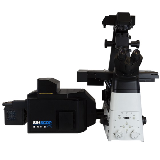 |
SIM Basic
Structured Illumination Microscope, SIM Basic, Image frame rate 13fps@1024x1024, Resolution ~100nm, image depth <50um, No. of Laser - 4, Wavelength choice 405/445/470/520/528/555/640nm, sCMOS Camera detectors, Inverted or upright microscope, XYZ motorized stages, Motorized filter electronics system, high image contrast, upgrade to SpinDisk SIM 100nm resolution
|
|
6-10 Weeks |
Request for quote |
|
|
|
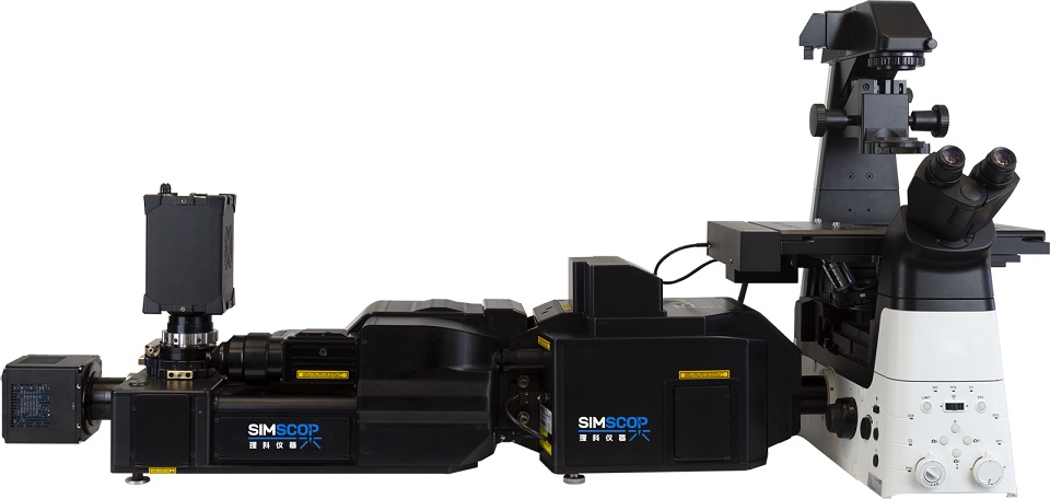 |
SpinDisk SIM
Structured Illumination Microscope, SIM Basic, Image frame rate 13fps@1024x1024, Resolution ~100nm, image depth <50um, No. of Laser - 4, Wavelength choice 405/445/470/520/528/555/640nm, sCMOS Camera detectors, Inverted or upright microscope, XYZ motorized stages, Motorized filter electronics system, high image contrast, upgrade to SpinDisk SIM 100nm resolution
|
|
6-10 Weeks |
Request for quote |
|
Accessories
| Compare |
Model |
|
Drawings & Specs |
Availability |
Reference Price
(USD) |
|