Spinning Disk Confocal Microscope
| Compare |
Model |
|
Drawings & Specs |
Availability |
Reference Price
(USD) |
|
|
|
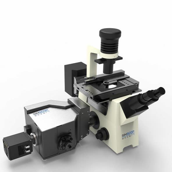 |
SpinDisk Basic Spinning Disk Confocal Microscope
Spinning Disk Confocal Microscope, SpinDisk Basic Series, Image frame rate 100fps@2048x2048, Resolution ~230nm, image depth <200um, No. of Laser - 4, Wavelength choice 405/445/488/525/561/640nm, sCMOS Camera detectors, Inverted or upright microscope, XYZ motorized stages, high image contrast, customized option
|
|
6-10 Weeks |
Request for quote |
|
|
|
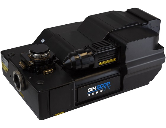 |
SpinDisk Advance
Spinning Disk Confocal Microscope, SpinDisk Advance Series, Image frame rate 100fps@2048x2048, Resolution ~230nm, image depth <200um, No. of Laser - 4, Wavelength choice 405/445/470/520/528/555/640nm, Dual sCMOS Camera detectors (single sCOMS optional), Inverted or upright microscope, XYZ motorized stages, Motorized filter electronics system, high image contrast, upgrade to SpinDisk SIM 100nm resolution, customized option
|
|
6-10 Weeks |
Request for quote |
|
SpinDisk Advance - Parameter
SpinDisk Basic Spinning Disk Confocal Microscope - Parameter
SpinDisk Advance - Download
SpinDisk Basic Spinning Disk Confocal Microscope - Download
Accessories
| Compare |
Model |
|
Drawings & Specs |
Availability |
Reference Price
(USD) |
|